Pancreatitis
The pancreas is located in the front part of the abdominal cavity. It is a boomerang-shaped organ made up of a right and a left lobe and a central part. The right lobe lies close to the descending duodenum, i.e. the first part of the small intestine, and this part produces most of the digestive enzymes. The left lobe lies between the stomach and the large intestine and secretes in particular enzymes that affect blood sugar balance.
The pancreas has several important functions in the body. It is divided into two parts: the exocrine and the endocrine part. The endocrine part produces, among other things, insulin in the so-called islets of Langerhans. Insulin moves glucose, i.e. sugar, obtained from food into use by the body, such as the muscles and the brain. The exocrine part mainly produces enzymes involved in digestion, such as lipase (phospholipase A).
Many mechanisms normally protect the pancreas from autodigestion. This means that the digestive enzymes cannot destroy pancreatic tissue, but only start working once they have reached the small intestine.

Pancreatitis
The actual cause of pancreatitis often remains a mystery. However, there are so-called triggering factors that have been found or are assumed to be causally linked to the development of pancreatitis.
These include:
- a diet low in protein and high in fat
- obesity
- certain drugs (suspects include azathioprine, oestrogens, tetracyclines, furosemide, potassium bromide, sulphonamides, etc.)
- backflow of intestinal contents (duodenal fluid reflux): e.g. vomiting or abnormal intestinal motility
- trauma or lack of oxygen affecting the pancreas (shock, severe anaemia, low blood pressure)
- infectious factors (in dogs, babesiosis)
Pancreatitis can be either acute or chronic. Acute and chronic pancreatitis cannot be fully distinguished on clinical symptoms alone; however, acute pancreatitis often has more severe symptoms and chronic pancreatitis milder ones. The changes in the pancreas caused by acute pancreatitis are reversible if the cause of the inflammation is removed. In chronic pancreatitis, irreversible changes have already taken place in the structure of the pancreas, such as normal tissue being replaced by connective tissue (fibrosis) and normal tissue wasting away (atrophy). Both forms of inflammation (acute and chronic) can be mild or severe, even fatal.
Symptoms of pancreatitis
Symptoms vary greatly depending on the severity of the disease. Mild cases may even have no symptoms. In the most severe cases, there may be loss of appetite, vomiting, weakness, abdominal pain, dehydration and diarrhoea.
A dog of any age or breed can get pancreatitis, regardless of sex. However, it is more common in middle-aged and older dogs. Overweight dogs are also in the risk group. The more severe form of pancreatitis occurs more often in males and spayed females.
Diagnosing pancreatitis
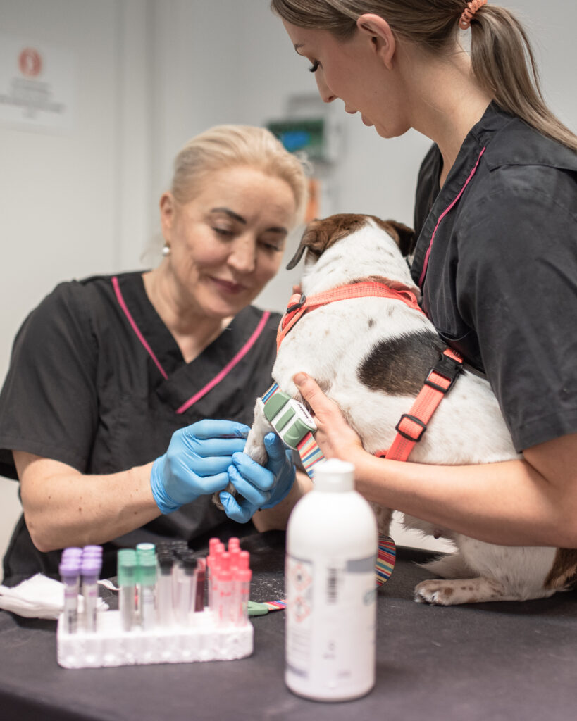
Blood test:
In pancreatitis patients, the blood count often shows an increase in total white blood cells (leucocytosis) and a so-called left shift (an increase in immature forms of white blood cells). Dehydration can raise the haematocrit (Hct), but anaemia may also occur in some patients. Azotaemia, i.e. raised urea in the blood, often occurs in connection with dehydration. Creatinine may also be elevated. Liver enzymes are often elevated because of ischaemia, i.e. lack of oxygen, in the liver and/or the burden caused by toxic substances produced by the pancreas. There may be more bilirubin in the blood than normal. Blood sugar is also often elevated. Some dogs may remain diabetic after pancreatitis. Hypocalcaemia, i.e. low calcium, occurs in pancreatitis patients and can cause mild muscle stiffness in some. Hyperlipaemia in the blood is common, even if the patient has not just eaten.
The pancreatic enzymes amylase and lipase are often elevated, but these are very non-specific values: lipase, for example, may also be elevated in gastrointestinal inflammation. In addition to the pancreas, lipase is also secreted from the first part of the small intestine.
cPLI (canine pancreas-specific lipase) is a value measured from the blood that measures lipase secreted by the pancreas. cPLI is the most sensitive test for pancreatitis, though it too is not infallible. According to studies, 81.8% of dogs with acute pancreatitis have elevated cPLI.
X-ray and ultrasound examination:
On an X-ray of the front of the abdominal cavity, taken with the dog on its back, on the right-hand side, there is typically increased radiopacity and loss of detail, and a gas-filled first part of the small intestine in an abnormal position. These changes are not always present despite pancreatitis.
Abdominal ultrasound is a very specific and diagnostic examination method in dogs. However, the examination requires a sufficiently high-quality ultrasound machine and an experienced ultrasonographer. Ultrasound shows local hyperechogenicity, especially in the chronic form, due to increased connective tissue. Reduced echogenicity, on the other hand, indicates necrosis in the pancreatic area. Increased echogenicity around the pancreas due to local peritonitis (inflammation of the abdominal lining) is also often seen.
Biopsy of the pancreas:
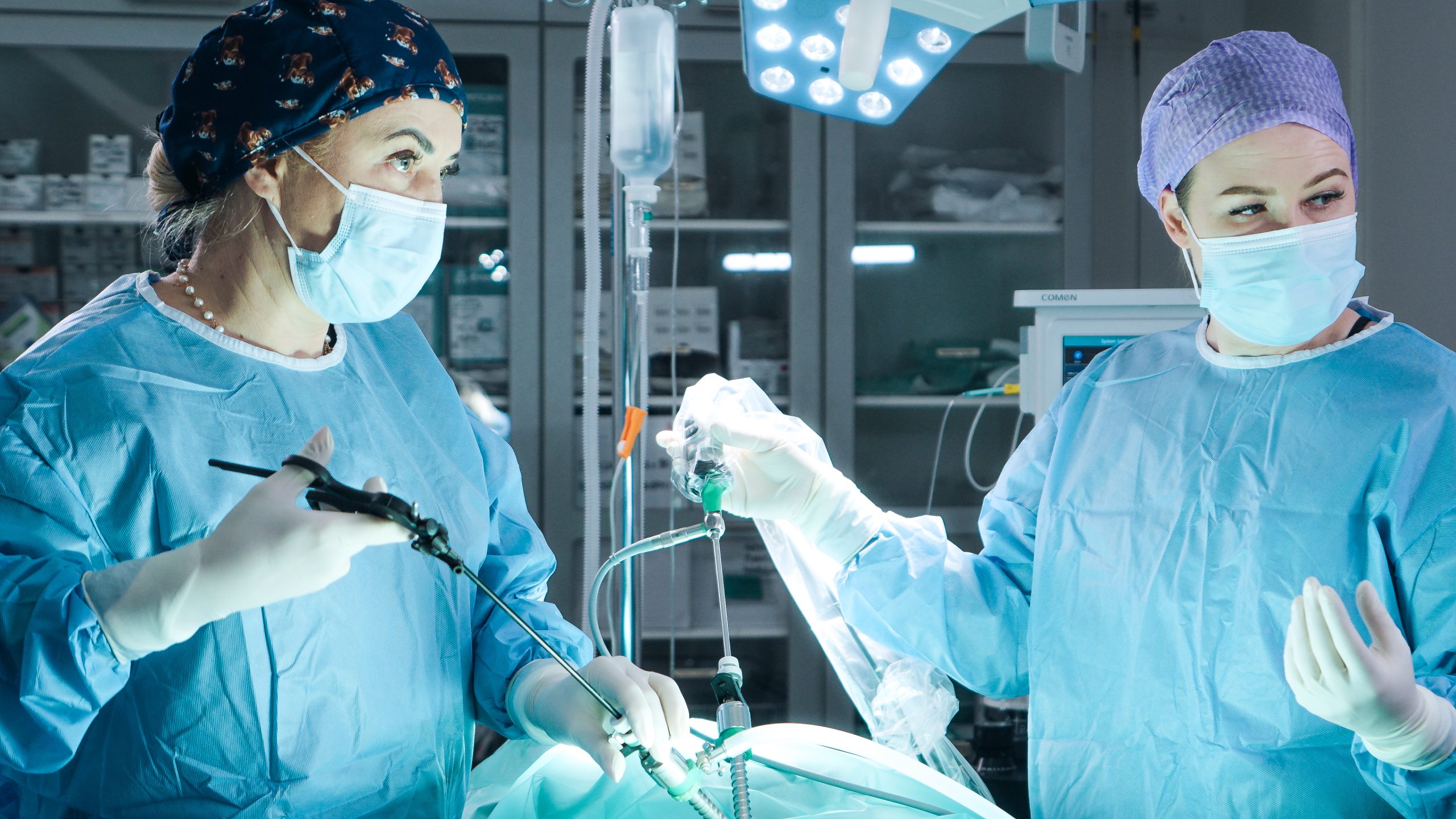
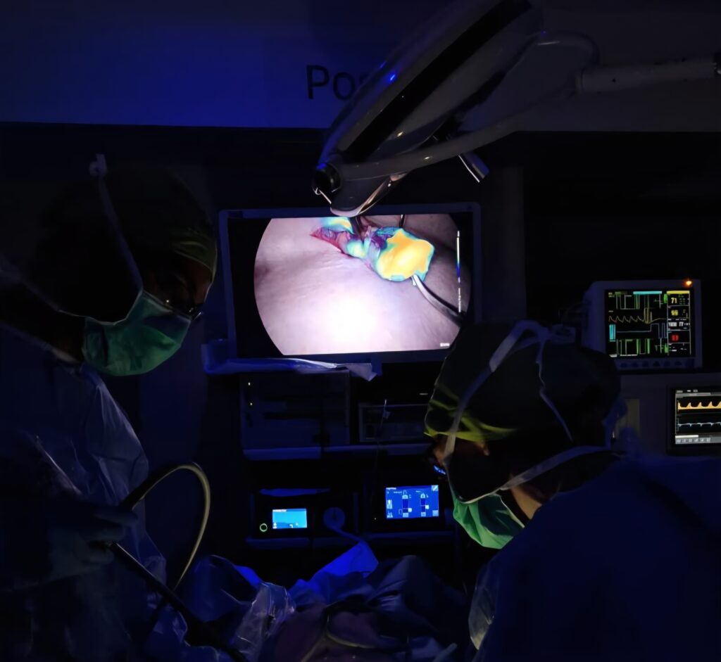
Taking a biopsy (or biopsies) of the pancreas is considered the best diagnostic method. However, the method is complicated by, among other things, the risks of anaesthetic complications in the acute phase of the disease and the “patchy” occurrence of the inflammation. Biopsies can be taken either traditionally by laparotomy, i.e. opening the abdomen, or by laparoscopy, i.e. endoscopically through the abdominal wall.

Treatment
The cornerstone of treatment is maintaining fluid and electrolyte balance with fluid therapy. Pain relief is also important, even if there are no clear signs of pain. Opioid painkillers are preferred for pain relief, because anti-inflammatory painkillers can cause serious side effects in a dehydrated, seriously ill patient.
The pancreas is allowed to “rest” by keeping the patient fasted until the nausea has stopped. Not even water is given by mouth; instead, fluids are given by drip.
In the past it was recommended that dogs be fasted for even long periods, but according to current knowledge a long fast can damage the intestinal villi, and so the intestine should be “fed” as soon as it is safe for the pancreas. Sometimes food can also be temporarily directed through a feeding tube past the pancreas (jejunal tube). Once water stays down, food is started in small portions. The food should be low in fat and protein but high in carbohydrates (rice, pasta, potato or ready-made intestinal prescription diets).
If a drug is suspected of triggering the disease, the medication must be stopped or replaced with another.
Mild cases can also be self-limiting, while more severe cases need hospital-level intensive care.
If abdominal pain occurs during the recovery phase, adding pancreatic enzymes to the food can be tried.
Prognosis
Pancreatitis is a disease whose course is hard to predict and which varies in severity. Severe forms in which other organs are also affected (DIC, SIRS, ARDS) have a poor prognosis. On the other hand, some individuals can survive with the right intensive care started early enough and make a full recovery. Most uncomplicated cases recover and do well, as long as fatty food/treats can be avoided in the future.